Your vision is important, so your eyes deserve to be looked after by trusted experts. Here at Brown Opticians we invest in the very latest consulting room and dispensing technology which allows us to go the extra mile for our patients. If you care about your eyes you’ll be reassured that we don’t rush appointments. We pride ourselves in always conducting a very thorough eye examniation to ensure our patients can make the most of their vision. Eye exams are crucial, no matter what your age or how healthy you feel.
During our comprehensive eye exam, our opticians will do much more than just determine your prescription for glasses or contact lenses. We will also check for common eye diseases and evaluate your eyes as an indicator of your overall health. Early diagnosis and treatment of eye and vision problems can help prevent vision loss, and other general health conditions can be referred to your GP or hospital.
Our eye examinations usually last about 30 minutes but we will advise you if you require any extra specific tests and how long these may take.
We have a general routine we perform for every patient, but as we like to treat every patient as an individual and our optician will tailor the examination similarly, which guarantees you a thorough examination executed smoothly, keeping you as relaxed as possible.
As well as including all tests to check for your sight and to see if any vision correction is required (e.g. spectacles or contact lenses) we also check for any potential eye disease like Glaucoma, Cataract, Macular degeneration to name a few.
Our optometrist will always explain the results of the various assessments and you will be given every opportunity to ask any questions you may have. The recommendations given at the end of the eye examination by the optician will take into account factors such as your working environment, lifestyle and any leisure pursuits you have, so we can meet your visual needs in the best possible way
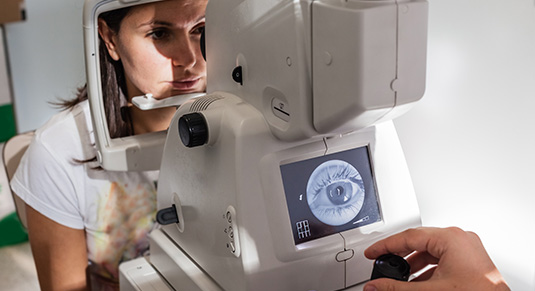
All eye examinations (subject to NHS recall guidelines) are funded by NHS Scotland. The NHS requires us to do retinal photographs for patients over 60, but at Brown Opticians we do this for everybody, free of charge. This extra test allows us to keep a permanent record of the back of your eye which means we can also monitor changes in your eyes between your visits to us.
Our equipment includes a digital retinal camera which allows us to take photographs of the inside of your eyes so even small abnormalities can be detected, recorded and compared in future checks. Combining this with modern examination techniques we can ensure all is done to help prevent unnecessary deterioration to your sight and eye health.
Other tests we will carry out include binocular assessment (how well the eyes work together), peripheral visual field analysis and intraocular pressure measurement.
Myopia
In Myopia (short sightedness), the eye is longer than normal or the cornea is too steep, so that light rays focus in front of the retina. Near objects are clear, but distant objects appear blurred. For the most part, this is an inconvenience considering how frustrating it can be to be dependant on contact lenses or spectacles.
In addition, eyes with a high degree of Myopia are at an increased risk of developing a serious condition like retinal detachment or glaucoma.
Hypermetropia
Hypermetropia (long sightedness), the eye is shorter than normal or the cornea is too flat, so that light rays focus behind the focused on clearly by the retina. Someone with hypermetropic eyes may find their vision is blurred when looking at objects near to them, and for vision to be clearer when looking at far away objects.
Placing a plus powered (convex) lens in front of a hypermetropic eye allows the image to be moved forward allowing for correct focus on the retina. A degree of long-sightedness is common in many people, although this only presents a problem when our ability to see is significantly affected or where headaches and eye strain are common.
Presbyopia
When we are young, the lens in the eye can change its shape allowing us to focus on near objects. After the age of 40, the lens becomes noticeably more rigid and reading at close range becomes increasingly difficult. This condition is called presbyopia and is a normal part of ageing. Presbyopia is usually first noticed by difficulties reading in low light. Often, you may find it will take longer for eyes to refocus from reading to distance and from distance to reading.
Spectacles may be required to give additional focusing power to the eye as reading proves more problematic. The distance of reading dictates which power you would require. For example, looking at a computer screen will require a different power for reading a book. We will ask you about your lifestyle and take this into account when prescribing your reading addition to ensure clarity of vision for the required visual task.
Glaucoma
We look at the condition of the optic nerve in all patients, and if over 30, we also measure the internal pressure. In around 95% of cases there are no syptoms for this condition, and glaucoma is often referred to as “the silent thief of sight”. The earlier the detection, the better the prognosis.
More InformationDiabetes
This is the most common cause of blindness in the working population in the UK today.
Any diabetic should have their internal eye health checked annually using pupil dilation in conjunction with retinal camera photography. These images will be archived for future comparison.
The diabetic retina characteristically shows a progression of circumstances including different types of “exudates”, “haemorrhages”,
“cotton wool spots” and ultimately end-stage retinal detachment. Although, with good blood glucose regulation most diabetics can prevent significant eye damage.
Macular Degeneration
Age-related macular degeneration (ARMD) is the commonest cause of vision loss in people aged over 50 years old. The prevalence (number of new cases each year) increases with age.
It is caused by degeneration of the macula, the central and most sensitive part of the retina at the back of the eye. There are two main types of ARMD often termed ‘Dry ARMD’ and ‘Wet ARMD’. The dry form is more common, but the wet form is usually more sudden and devastating to the vision.
More InformationCataract
Cataracts are extremely common. In fact, the majority of those over 65 have some cataract development.
If you have been told you have cataracts, DO NOT be alarmed. A cataract simply refers to ‘opacity of the lens’ inside the eye. Looking through a cataract can be thought of as a little bit like looking through an old stained piece of glass – instead of a clear new sheet.
There are many different types of cataract. Not all cataracts cause symptoms. If a cataract causes no symptoms, it can usually be left alone. If symptoms such as blurred vision occur, then cataracts can be treated very successfully with surgery.
More InformationRetinal Detachment
There are three types of retinal detachments.
The most common form is where a break in the retina’s sensory layer causing fluid to seep underneath. This eventually causes a separation in the layers of the retina. Individuals who are particularly short sighted, with historic eye injuries or who have undergone eye surgery are most susceptible to this type of detachment. This is due to the thinner and more fragile retina in short sighted people.
The second most common type is due to increased traction on the retina by strands of scar or vitreous tissue which can ultimately pull the retina loose.
The third most common type occurs when small pockets of liquid form within a special gel (the virtreous) which usually lines the inside of the eye. Eventually, some of this fluid moves in between the gel and the retina, causing the vitreous to peel away from the retina. If a hole or tear develops in the retina, then there is an increased risk of there being a retinal detachment.
A detached retina can cause loss of vision, and requires a surgical operation to put the retina back in the right place. Thus, it is very important that you have your eye examined urgently on the onset of symptoms.
More InformationFloaters and Flashing Lights
Floaters are extremely common, and are sometimes associated with flashing lights in the eye, especially when they first appear. When they first appear, they normally affect one eye, but may affect both eyes at the same time.
In fact, they’re so common, that approximately two thirds of the population will have floaters by the time they are in their mid sixties! However, they can occur at any age.
More InformationDry Eyes
Dry eyes occur when the eyes either don’t make enough tears, or the quality of the tears produced is reduced, which means the tears can evaporate rapidly from the surface of the eye, allowing the eye to dry.
Often, the reduced tear quality is a result of blockage or inflammation of the oil glands within the lid margin. When the surface of the eyes dry out, the eyes become inflamed. They appear red, and the whites of the eyes can appear to be pink and swollen. Normally, the eyes become very irritable.
It is more common in women than men, and is found most commonly in the over 60s age group. However, it can happen at any age.
The symptoms can be extremely variable, causing anything from mild irritation to severe discomfort. Symptoms include:
• Foreign body sensation/feels like something is in the eyes
• Eyes feel ‘gritty’ – often worse in the mornings
• Blurred vision
• Burning sensation in eyes
• Irritable eyelids
• Light sensitivity
• Redness of the whites of the eyes
• Painful eyes
• Excessive watering
There is no absolute ‘cure’ for dry eye syndrome. However, most people can get significant relief from symptoms using a variety of treatments and measures.
Regular lubrication in the form of gels or drops can help keep the surface of the eyes wet, and thus reduce symptoms. Often, this is combined with lid margin hygiene. There are a wide range of eye drops available – consult our optometrist for advice on which one to use.
More InformationBlepharitis
Blepharitis refers to inflammation of the eyelid margins. There are two broad categories of the condition – Anterior and Posterior Blepharitis.
Blepharitis is very common indeed. People of any age can be sufferers, but it is more common in older people over the age of 50.
It is not something ‘caught’ or inherited. The reason for some people developing blepharitis is poorly understood.
Blepharitis is normally diagnosed easily during your eye examination with a look at your eyelids through the microscope. Simple visualisation of the lid margin along with the history is how the diagnosis is normally made.
More Information- 20 million British people risk avoidable sight loss because they fail to have regular sight tests.
- 1 in 10 British adults have NEVER had an eye examination.
- 85% of us admit to having problems with our vision.
- More than 30 million Britons are entitled to FREE eyecare (sight tests and / or optical vouchers to cover the cost of any vision correction required) paid for by the NHS.
- Everyone should have an eye examination once every two years unless advised otherwise by their optometrist. The Eye Care Trust recommends children aged under 9 and people aged 70 and over have annual eye examinations.
- It’s estimated that 1 in 5 children has an undetected problem with their vision.
- 50% of us think an NHS sight test costs £20 or more, despite it being free!
- A quarter of over 60 year olds say the quality of their vision affects their daily routine. Source of information: The Eyecare Trust.